YouTubeアイコン
ページトップへ

Keratoconus

What is Keratoconus?
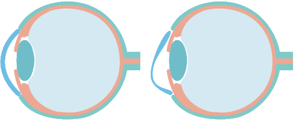
Keratoconus is a disorder of the eye which results in progressive thinning of cornea.
Normal cornea and keratoconus

Normal cornea

Keratoconus

Keratoconus is a disorder in which the shape of the cornea changes, myopia and irregular astigmatism intensitfy, and as it progresses, it cannot be corrected even with glasses or contact lenses, and corneal transplantation is required.

The cause is unknown and it is a congenital, progressive disease. Eye-rubbing irritation (corneal abrasion) such as atopy and allergic disease is said to be an exacerbating factor, but it may develop without such an incentive.

There are no subjective symptoms in the initial stages. As the disease progressive, astigmatism increases, but good corrected visual acuity can be achieved with glasses or contact lenses. Therefore, it is rarely detected in the early stages in most case and is generally detected after detailed analysis of corneal shape by means of testing such as for refractive surgery. Keratoconus develops in most people during adolescence in the second and third decades of their life, then progress, and become less likely to progress after the age of 40. Rarely, it is discovered after the age of 30 and progresses after the age of 50. It is said to affect 1 in 1,000 people, and it is not a rare disease.

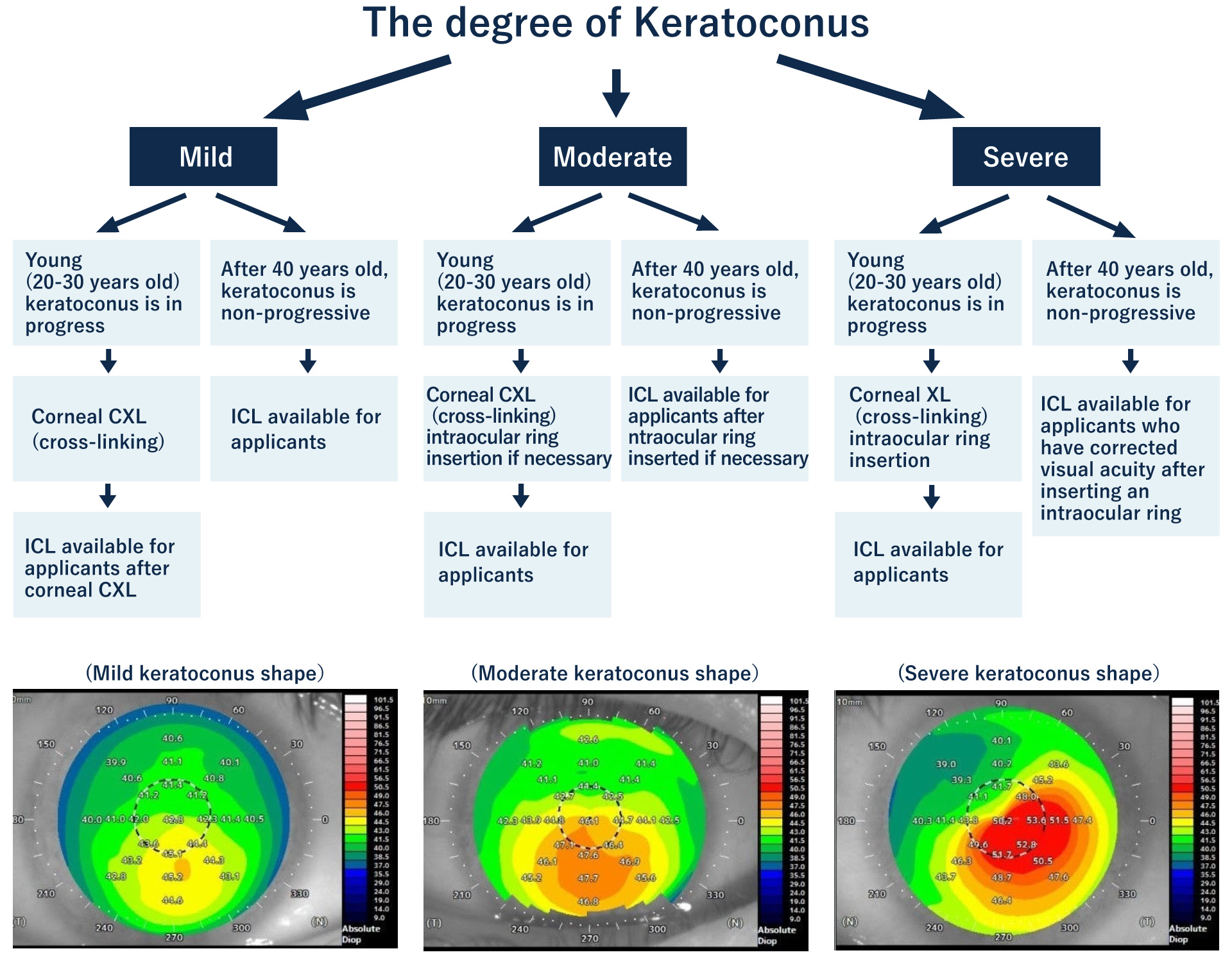
The treatment of keratoconus is different according to the development of cornea.

At Eye Clinic Tokyo, combining the treatment for keratoconus and ICL is suggested to greatly reduce the degree of myopia and astigmatism in those with stable corrected vision and thicker cornea than specified.

In February 2019, the Japanese Society of Ophthalmology revised the “guidelines for refractive orthodontic surgery (7th Edition)”, “previously considered to be contraindicated” for keratoconus cases in the treatment of ICL. Now the possibility of treatment has expanded. Kitazawa, who is in charge of the operation, is certified as an ICL expert instructor, with only ten people in Japan having this certification, and has rich experience with respect to providing patients with world-class treatment.

Keratoconus treatment and
ICL flow chart

Keratoconus treatment and ICL flow chart Keratoconus treatment and ICL flow chart

Note) Because there is a risk of progression in keratoconus around the age of 20, it is recommended to first suppress the progression with corneal collagen cross-linking and then undergo ICL surgery after observing the progress of keratoconus and myopia.

    Degrees of keratoconus

  • ■Mild

    • ①There is slight astigmatism and it cannot be detected by ordinary ophthalmic examination.
    • ②Good corrected visual acuity can be achieved with glasses or contact lenses.
    • ③Keratoconus can be diagnosed for the first time with a corneal shape analyzer.

    ■Moderate

    • ①Increasing astigmatism that can progress to oblique astigmatism.
    • ②Glasses and soft contact lenses can no longer correct vision acuity; therefore, hard contact lenses should be used to correct vision acuity.
    • ③It is sometimes said that keratoconus is suspected in general ophthalmic examinations.

    ■Severe

    • ①Increased astigmatism, visual acuity cannot be corrected using normal glasses and soft contact lenses.
    • ②Good corrected visual acuity can be achieved with hard contact lenses, but the lenses can easily move.
    • ③visual acuity is corrected with special hard contact lenses (custom-made) for keratoconus.
    • ④Wearing a hard contact lens on top of a disposable soft contact lens or Eye Brid TM, or wearing lens having hard contents and soft contact lenses around it can improve the discomfort felt while wearing contact lenses.

    Treatment for Keratoconus

  • ■Corneal Cross-Linking

    • ①Because the main purpose is to suppress the progression of keratoconus, this treatment is indicated for young people with keratoconus or those at high risk of keratoconus.
    • ②In recent years, it is suggested that early treatment should be started at the age of 13 - 20 years for keratoconus formation.
    • ③Corneal cross-linking (especially topography-guide cross-linking) can reduce myopia and astigmatism in addition to suppressing the progression of keratoconus.
    • ④The biggest drawbacks of treatment are pain for approximately 2 days after surgery and it takes approximately 1 week for the eyesight to improve.
    • ⑤You should be careful if you have atopic or allergic constitution and are used to rubbing eyes because keratoconus may develop even after corneal collagen cross-linking surgery.

    ■Intrastromal Corneal Ring Segment Implantation (ICRS) Implantation

    • ①Because the main purpose is to improve the irregular shape of keratoconus and make it easier to obtain corrected vision, it is suitable for those who have advanced keratoconus to some extent and for those who do not have clear vision with eyeglasses or contact lenses.
    • ②In young people, corneal collagen cross-linking may be performed before or simultaneously with intracorneal ring segment (ICRS) implantation.
    • ③Although astigmatism and myopia can be reduced through the ICRS implantation, it does not mean that such people can live with naked eyes.
    • ④The biggest drawbacks of treatment are that it takes approximately a month for postoperative vision to stabilize and night vision is blurry.
    • ⑤Even with ICRS surgery, those who have the habit of rubbing the eyes may also develop keratoconus; therefore, it is necessary to pay attention to not rubbing the eyes.

    ■ICL

    • ①ICL has no effect on improving keratoconus.
    • ②If you have early keratoconus or suspected keratoconus and are unlikely to develop into keratoconus with age, then it’s possible to have ICL surgery.
    • ③Young people with keratoconus are treated with appropriate treatments such as corneal cross-linking or ICRS implantation. ICL surgery is available if keratoconus progression is suppressed.
    • ④The visual acuity after ICL surgery is lower for keratoconus than that for normal myopia and astigmatism.
    • ⑤The expected corrected visual acuity after ICL surgery depends largely on keratoconus. ICL is as one of the highest visual acuity that can be corrected with glasses, but it may not be as good as the corrected vision with glasses.
    • ⑥Although it is possible to live with the naked eye after the ICL operation, vision equivalent to that obtained on using hard contact lens and special contact lens for the conical cornea cannot be expected.

    Specific examples of keratoconus treatment
    and ICL surgery

  • Mild cases of keratoconus

    Case 39-year-old male right eye

    Preoperative:
    Vd=0.06(1.5X-6.25D cyl-1.25D Ax170)
    Naked eyesight 0.06
    Corrected vision 1.5
    Myopia power -6.25D
    astigmatism power -1.25D
    39-year-old male; Myopia and the initial keratoconus did not progress and the corrected visual acuity was good, only ICL surgery was performed.

    6 months after ICL:
    Vd=1.5(n.c.)
    Naked eyesight 1.5
    Corrected vision 1.5

  • Moderate case of keratoconus

    Case 44-year-old male right eye

    Preoperative:
    Vd=0.07(1.2X-8.50D cyl-2.25D Ax100)
    Naked eyesight 0.07
    Corrected vision 1.2
    Myopia power -8.50
    astigmatism power -2.25D
    moderate keratoconus did not progress at the age of 44 and the corrected visual acuity was good, only ICL surgery was performed.

    3 months after ICL:
    Vd=1.2(1.5XICLX-0.50D cyl-0.75D)
    Naked eyesight 1.2
    Corrected vision 1.5

  • Moderate case of keratoconus

    Case 31-year-old male right eye

    Preoperative:
    Vd=0.6(1.2X-0.25D cyl-3.00D Ax5°)
    Naked eyesight 0.6
    Corrected vision 1.2
    Myopia power -0.25
    astigmatism power -3.00D
    Moderate keratoconus was also advanced at the age of 31 years, and corneal cross-linking was performed first, and ICL surgery was performed 6 months later.

    1 month after ICL:
    Vd=1.0(1.2XICL cyl-0.75D Ax5°)
    Naked eyesight 1.0
    Corrected vision 1.2

  • Severe case of keratoconus

    Case 46-year-old male right eye

    Preoperative:
    Vd=0.1(0.8X-2.50D cyl-5.25D Ax120)
    Naked eyesight 0.1
    Corrected vision 0.8
    Myopia power -2.50D
    astigmatism power -5.25D
    At the age of 46, keratoconus was progressing and corrected visual acuity was also impaired, so corneal cross-linking and intraocular ring surgery were performed at the same time, and ICL surgery was performed 6 months after the surgery.

    6 months after ICL:
    Vd=0.5(1.5X-1.25D cyl-0.50D Ax45°)
    Naked eyesight 0.5
    Corrected vision 1.5
    The corneal shape improved, the astigmatic power decreased to -0.50D, and the corrected visual acuity also improved to 1.5.

If you are using a personal computer, please read this QR code on your smartphone and use it.

If you want to inquiry via LINE, please click here.

Inquiry LINE:@eyeclinic-tokyo

@eyeclinic-tokyo

If you want to contact us via WeChat, please contact here.

Inquiry WeChatID:eyeclinictokyo

If you want to contact us via Phone, please click here.

080-8496-2829

Reception hours: 10:00-18:00 (excluding Thursdays)